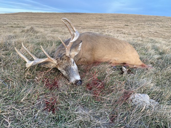


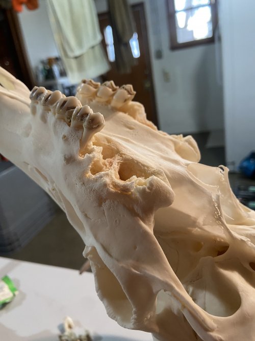
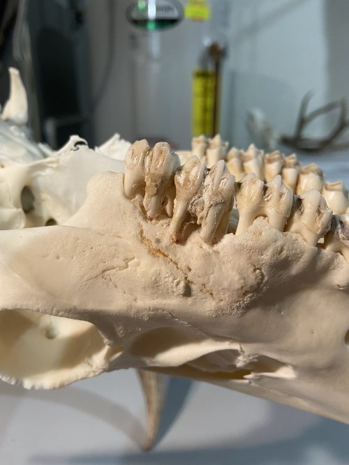
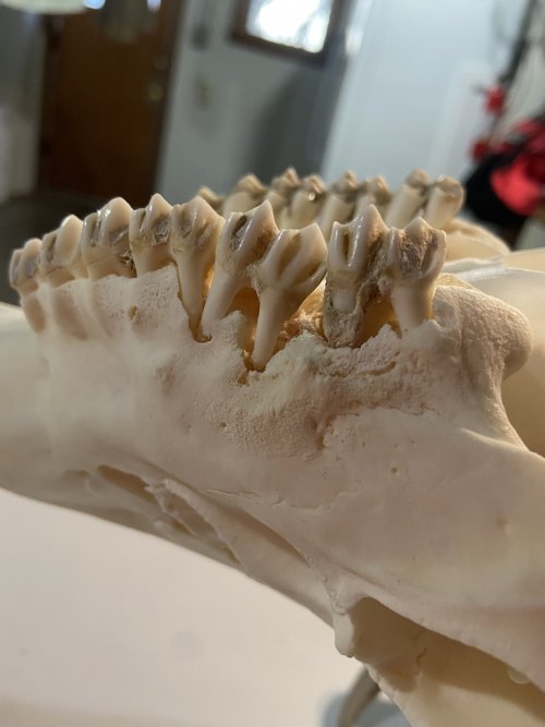
I shot this buck in North Central SD a couple weeks ago, and while skinning the skull to euro it I discovered an awful stench in the jaw area that was associated with a large infected pus pocket on one side and a smaller on the other, but not as bad.
I figured it was due to fighting and didn’t think much other than it was gross and sucked to take care of.
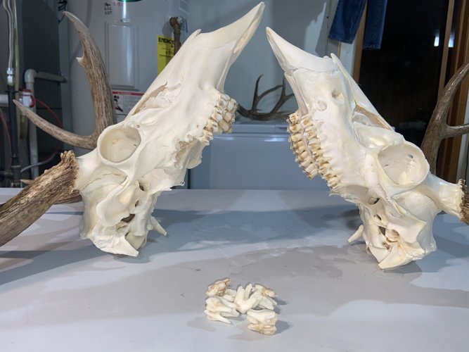
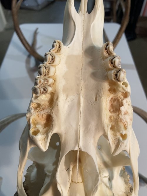
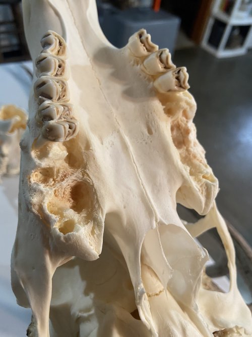
After an initial few hours in a simmer, I pulled it out to do some fleshing and teeth started falling into the water. Turns out the skull where the pus pockets were is pretty deteriorated and honeycombed, almost.
Just wondering if anyone has any idea what this might be from? It was very localized to the jaw area and the rest of the meat and carcass was perfect, besides a fracture in his maxilla near his nasal cavity, which I’m almost positive would be from fighting.
He was a 4x4 whitetail, probably 3-1/2 years old based on rack and body size.
I figured it was due to fighting and didn’t think much other than it was gross and sucked to take care of.
After an initial few hours in a simmer, I pulled it out to do some fleshing and teeth started falling into the water. Turns out the skull where the pus pockets were is pretty deteriorated and honeycombed, almost.
Just wondering if anyone has any idea what this might be from? It was very localized to the jaw area and the rest of the meat and carcass was perfect, besides a fracture in his maxilla near his nasal cavity, which I’m almost positive would be from fighting.
He was a 4x4 whitetail, probably 3-1/2 years old based on rack and body size.
Attachments
-
 03B136C0-C444-477B-8132-F02D8EF6319C.jpeg2.2 MB · Views: 78
 -
 EAD62733-3166-4A32-8EDD-7B19B7E19756.jpeg1.3 MB · Views: 75
 -
 2C9BA52E-9758-48F3-8513-68E426F355B6.jpeg1.4 MB · Views: 62
 -
 716B5C3E-1148-4D19-83D9-34EA98E16901.jpeg1.3 MB · Views: 63
 -
 58D0DB52-3444-4F79-9E4E-DEA7ED06F17B.jpeg1.6 MB · Views: 64
 -
 91C7A6EA-BF62-4273-9584-A3BBC5EE5F31.jpeg1.3 MB · Views: 55
 -
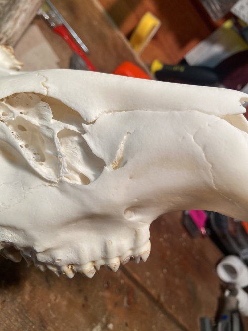 image.jpg1.6 MB · Views: 72
Last edited:
")